Osteoporosis is a silent disease characterized by decreased bone mass and deteriorated bone quality leading to increased fracture risk. Osteoporotic fractures are associated with high rates of disability, loss of independence, reduced quality of life for patients and caregivers, and high costs to individuals and healthcare systems. Nevertheless osteoporotic fractures are often overlooked and bone mineral density screening rates are low.

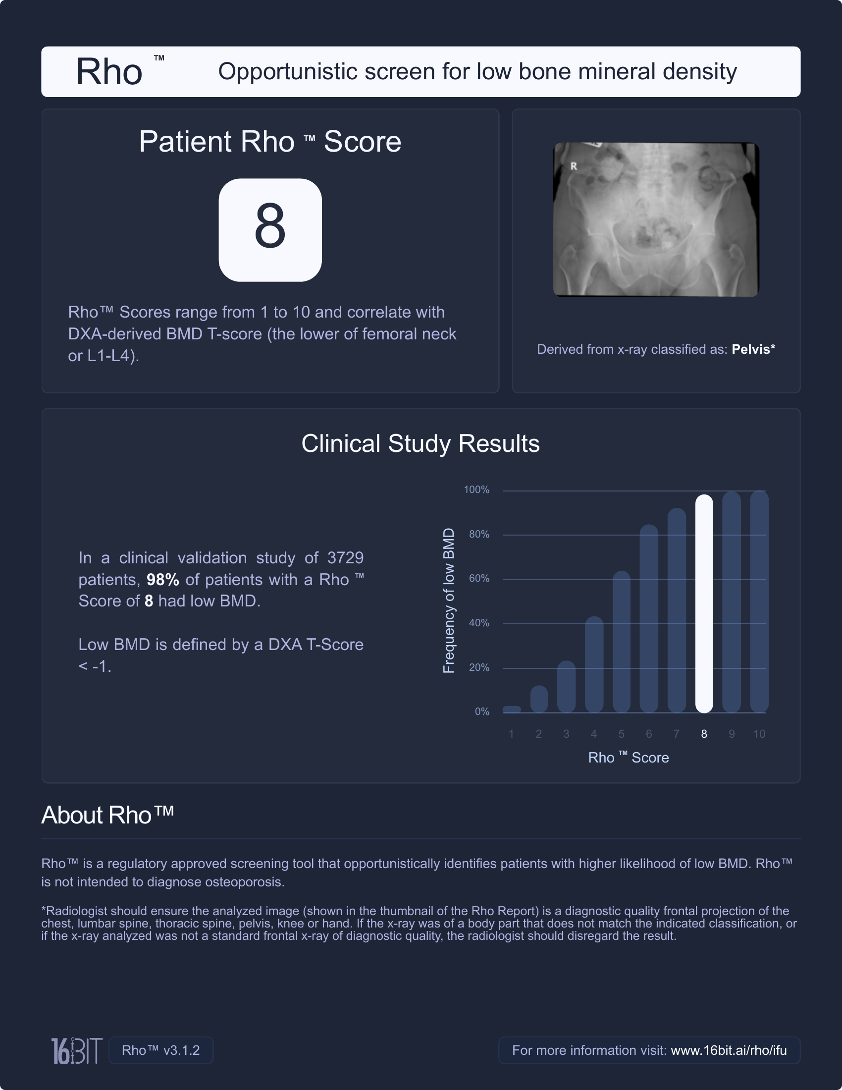
For patients aged 50 and older, a Rho™ Score and Rho™ Report are automatically generated when suitable X-rays are taken (e.g., spine, chest, pelvis, knee, or hand). The Rho™ Score ranges from 1 to 10, indicating the likelihood of low bone mineral density.
An individually defined threshold determines when Rho™ Reports are automatically sent to PACS. Radiologists can include this information as an incidental finding in their report, providing the referring clinician with a basis for further evaluation, such as a DXA scan.
This enables fracture risk assessment to occur earlier than in current practice, supporting the timely initiation of preventive and therapeutic measures.
assess bone health whenever a suitable X-ray is taken
the Rho score indicates a T-Score < -1
assists radiology reporting efficiently
Rho-positive patients confirmed by DXA
AUC - Excellent risk detection performance across anatomical regions
Images (X-ray/DXA datasets) were used to train Rho
Rho™ is a medical device software application that interfaces with institutional Picture Archiving and Communications Systems (PACS) to estimate the likelihood that a patient over the age of 50 undergoing x-ray has low bone mineral density (BMD).
Eligible x-rays are frontal projections of:
Rho™ generates a score from 1 to 10 to indicate the likelihood of low bone mineral density (DXA T‑Score < ‑1), with adaptable thresholds for flagging patients as “positive” and a graphical summary of findings for easy interpretation.
Rho™ is a software application intended for use with x-ray images from radiological exams.
Rho provides a Rho Score risk-of-low-BMD category to aid health care professionals in the assessment of risk of low BMD. Rho produces adjunctive information. It is not a diagnostic aid.
When x-rays are being acquired for any clinical indication, Rho is intended to take the opportunity to analyze those existing x-rays. The intended target is patients aged 50 years or older, only if (i) at least one of their x-rays is of one of the validated x-ray types that Rho can analyze, and (ii) Rho detects that the patient has not had a recent dual-energy x-ray absorptiometry (DXA) scan at the imaging centre. In the latter case, the patient’s bone health would already be known. Rho can analyze frontal radiographs of the lumbar spine, thoracic spine, chest, pelvis, knee, or hand. A customer site can choose the time period for checking for recent DXAs. For example, a site may choose that Rho will not analyze an x-ray if the patient has had a DXA in the last 3 years.
16BIT homepage. https://www.16bit.ai/
Bilbily, A., Syme, C. A., Adachi, J. D., Berger, C., Morin, S. N., Goltzman, D., & Cicero, M. D. (2024). Opportunistic screening of low bone mineral density from standard X-rays. Journal of the American College of Radiology, 21(4), 633–639. https://doi.org/10.1016/j.jacr.2023.07.024
International Osteoporosis Foundation (2026): Educational Hub. https://www.osteoporosis.foundation/educational-hub#main


ImageBiopsy AI software is highly accurate and efficient within our PACS system, which provides valuable information on the status of the knee along the continuum of chondrosis to arthrosis.


The integration of the AI solutions by ImageBiopsy Lab into our RIS and PACS is easy and well done. It is fun to work with and the clarity of the visualized report is an ideal support for our patient consultation.

AI-based solutions reduce the amount of work and the findings become more accurate. An objective value is given which can be used both for monitoring and forecasting the progress. We offer something that others don’t have.

Exact diagnosis and reproducible follow-up exams are indispensable for a successful osteoarthritis therapy. Software-based methods can assist the physician in the therapy management and adjustment process.


